Our research

Cell polarity is the process of creating asymmetry. It is essential for normal cell function and for several key developmental processes, such as cell migration, axis determination and asymmetric stem cell divisions. There are many different flavours or axes of polarity. Epithelial cells are polarized along an apico-basal axis, and this polarisation is required for tissue integrity, whereas loss of polarity is a critical step in the formation of tumours. We are using Drosophila and mouse intestinal organoids to analyse how polarity arises and how cortical polarity factors regulate other polarised aspects of cell behaviour.

temporary image

The canonical epithelial polarity system

Hélène

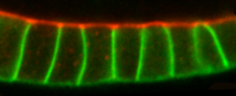
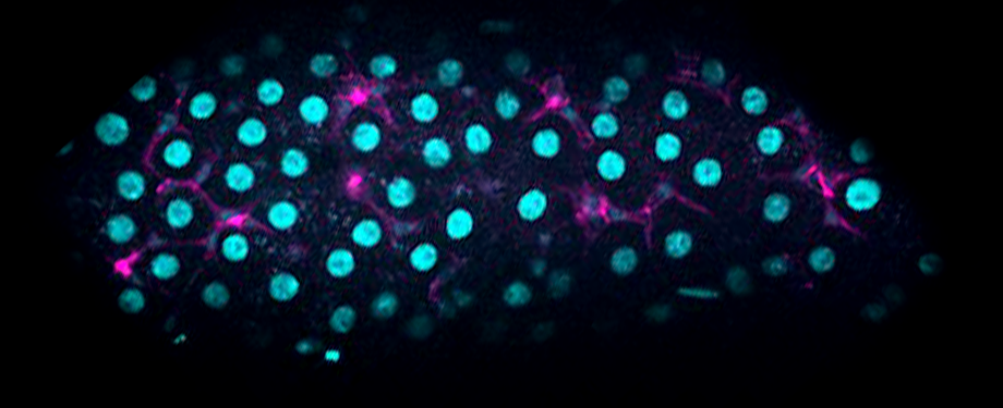
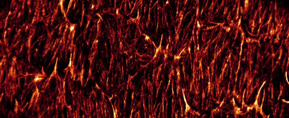
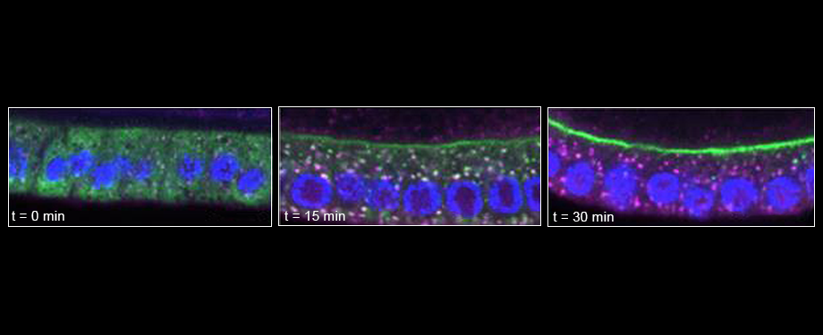
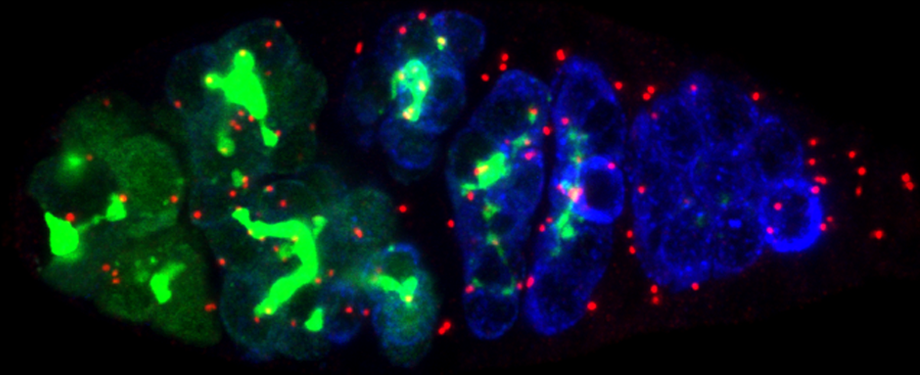
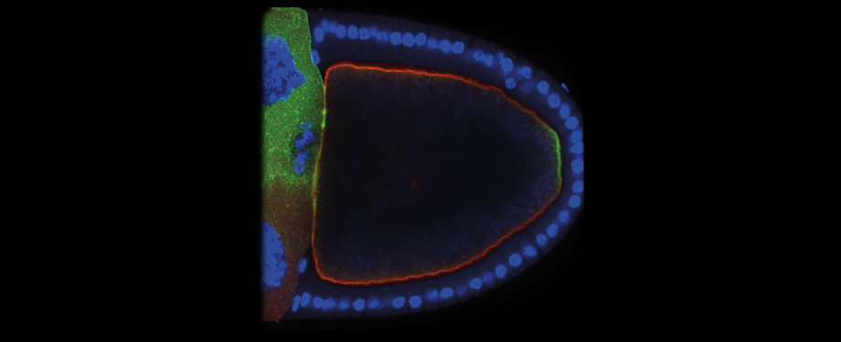
Epithelial cells form sheets that act as barriers which line our organs. To form these sheets the cells must be polarised along their apico-basal axis. Studies in Drosophila have identified a conserved set of polarity proteins responsible for establishing and maintaining epithelial polarity. The polarity proteins can be separated into apical factors (including Crumbs, Stardust, Par-6 and atypical protein kinase C [aPKC]), junctional (Bazooka/Par-3) and basolateral (including Scribble, Discs large and Lethal(2) giant larvae). In addition, Cdc42-GTP acts upstream of Par6/aPKC for apical domain formation. We are using proteomics, live cell imaging and mutant clone analysis to dissect the temporal relationships between these polarity proteins, as well as factors regulating Cdc42 activity.

temporary image

The alternative polarity system of the midgut

Jia | Joseph | Mihoko

Whilst all other epithelia in Drosophila are polarised by the canonical polarity programme the intestinal epithelial of the midgut is special. We have found that its polarisation is dependent on integrin adhesion and septate junction proteins rather than the canonical polarity factors such as Crumbs and Bazooka, which are not detectably expressed. There are a number of features that make the midgut tissue different, i) it is derived from the endoderm, whilst all other fly epithelial are ectodermal or mesodermal in origin ii) the occluding junctions are apical to the adherens junctions iii) enterocytes are polarised from basal to apical as they integrate into the epithelial layer. Together these features suggest that the midgut epithelium represents a better model for dissecting polarity pathways in vertebrates. To explore midgut polarity in more detail, we are undertaking large scale genetics screens and developing live imaging protocols.

temporary image

Super-resolution microscopy in tissues

George | Edward | Jaime| Jenny | Xiaoming

Many key biological processes happen below the resolution limit of conventional light (fluorescence) microscopes (less than 250 nm). This is known as the diffraction limit. Beginning in the early 1990’s, a number of techniques to circumvent the diffraction limit have been devised all with the target of multicolour imaging at the nanoscale in biological specimen. As these techniques have matured, their use in biological imaging has become more routine but limitations still exist particularly when working away from the cover glass in samples with high background. Our research is focused on pushing existing super-resolution methods into thicker specimen, specifically Drosophila egg chambers, in user friendly platforms accessible to non-experts. We work with two flavours of super-resolution: stimulated emission depletion (STED) and localization-based methods (STORM and DNA-PAINT) with an interest in 4Pi microscopy. You can find out more about what we do on our sub-group website.

temporary image

Polarised secretion

Jenny | Xixi

Membrane trafficking describes the movement of proteins and lipids around cells. Proteins and lipids must be accurately sorted between the organelles of the cell and the plasma membrane. In epithelial cells the trafficking itinerary is more complex because of the presence of distinct domains within the plasma membrane, traditionally split into apical and basolateral. Accurate trafficking to these domains is key to both the establishment and maintenance of polarity. Our research is focussed on how proteins move to specific domains of the plasma membrane in Drosophila epithelial cells. We use the RUSH system to retain our cargoes of interest in the ER and then are able to image a burst of trafficking through the secretory system. By studying this movement in wild type and mutant tissue we can gain insight into the different trafficking routes required to maintain the characteristic distribution of these proteins. We are collaborating with optical engineers and computer scientists to optimise both our imaging and image analysis.

temporary image

Polarised microtubule organisation

Dmitry

Microtubules are dynamic polymers which provide intracellular tracks for various motors to transport proteins, vesicles and RNAs around the cell. During cell polarisation, microtubules have to be re-arranged to direct cargo traffic to specific cellular compartments. However, how polarity factors organise microtubules into polarised networks is very poorly understood. Recently, we found that microtubules are organised by noncentrosomal microtubule organising centres (ncMTOC) in differentiated cells. ncMTOCs are distributed in the cell in a polarised fashion: they localise apically in epithelial cells and at the anterior-lateral cortex in the oocyte. Although ncMTOCs are the main source of microtubules in these cells, very little is known about their protein composition, how their activity is regulated and how their distribution in the cell is regulated by polarity cues. We use the Drosophila follicular epithelium, the oocyte and female germline cysts as model systems to find answers to these questions.

temporary image

Axis formation

Hélène

Correct body plan formation in Drosophila depends on the establishment of two axes of polarity in the developing oocyte, anterior-posterior and dorsal-ventral. Par-1 kinase is one of the master regulators of the anterior-posterior axis. It is recruited to the posterior cortex of the oocyte during mid-oogenesis and is required to polarise the microtubule cytoskeleton that directs the localisation of the axis determinants, oskar and bicoid mRNA. We are currently working on factors upstream of Par-1 that specify the A-P axis. The dorsal-ventral axis is set up when the nucleus migrates from the posterior of the oocyte to the dorsal/anterior corner. We are trying to identify the follicle cell signal(s) that regulate this migration.