
Our major focus is on how mitosis is controlled by cyclin-dependent kinases and ubiquitin-mediated proteolysis. We use real-time DIC and fluorescence microscopy to determine when and where proteins act and are destroyed.
We use proteins fused to Green Fluorescent Protein and its derivatives to assay protein dynamics, and as a read out of protein levels. By quantifying the fluorescence of our GFP-chimaeras we can assay the level of the protein. This gives us a live-cell assay for proteolysis. By this means we can determine when a protein begins to be destroyed, and thereby identify the event that triggers its destruction. (See Movie 1 as an example.)
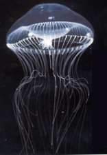
Aequoria victoria
Photo courtesy of Dr Claudia Mills (http://faculty.washington.edu/cemills/Aequorea.html)
We use homologous recombination to target GFP into the endogenous locus for cell cycle regulators. This allows us to follow the behaviour of the endogenous proteins in living cells, to measure molecule numbers and to derive accurate kinetics for protein destruction.
Recently we have been developing biosensors to detect protein kinase activity in living cells by Fluorescence Resonance Energy Transfer (FRET). (See Movie 5 as an example of a FRET assay.)
We have developed a biosensor specific for Cyclin B1-Cdk1 that allows us to measure the kinetics with which Cyclin B1-Cdk1 is activated as cells enter mitosis (see Gavet and Pines, 2010).
To determine the mechanisms underlying mitosis we are combining biochemistry with live cell imaging. We are analysing mitotic protein complexes by mass spectroscopy and reconstituting their activity in vitro (see Pagliuca et al., 2011).
We now intend to use our quantitative imaging and biochemistry to develop predictive models for cell cycle regulation.

Pablo Picasso: The Guitar Player (1910)